DOI: https://doi.org/10.37756/bk.21.3.3.1
Article type: Perspective
Received: February 2, 2021
Revised: February 15, 2021
Accepted: February 18, 2021
To cite this article: Tripathi P., et.al., BioMEMS for Life-Saving Biomedical Technologies, Biotechnol. kiosk, Vol 3, Issue 3, PP: 5-15 (2021); DOI: https://doi.org/10.37756/bk.21.3.3.1.
Abstract
Lately, there have been huge research interests in developing micro-electro-mechanical systems (MEMS) for various biological and medical applications. These bioMEMS based devices are considered instrumental to develop many life-saving biomedical technologies. To this end, a number of studies have focused on the developments of bioMEMS in the field of molecular biology, biotechnology, medicine, biochemical and material sciences and also in microsystems technology. The applications of bioMEMS are extensive that include diagnostic research, drug delivery, therapeutics, tissue engineering, biosensors and lab-on-a-chip systems for regenerative medicine, to name a few. Here, we present a perspective on the important breakthroughs in bioMEMS including the advances in microfabrication, monitoring and modulating cellular activities along with notable applications of bioMEMS in the modern healthcare sector.
Keywords: BioMEMS; Cardiovascular Disease; Microfabrication; Cell Culture; Therapeutics; Organ-on-a-chip
- Introduction
Last few decades have witnessed notable research efforts to develop highly controllable microscale fabrication techniques for applications in a wide range of biomaterials substrates. These efforts have subsequently led to the development of biological micro-electro-mechanical-systems ‘bioMEMS technology, which represents an emerging new area of biotechnology [1]. The field of bioMEMS technology is enabled by the combination of a number of technologies including electronics, fluidics, optics, sensors, and micro/nanotechnology. BioMEMS devices offer a vast array of life-saving biomedical technology applications. These include genomics and proteomics, early lab-on-a-chip devices for point-of-care (POC) diagnostics as well as clinical diagnostics, toxicity screening to artificial organ assist devices, implantable tissue constructs and also in-vitro tissue models for drug delivery systems. The design principles of BioMEMS fabrication techniques are based on low-cost, simplicity and ease of processability. These are the technology drivers of bioMEMS fabrication techniques. Researchers have leveraged these principles to build new and emerging technology platforms such as organ-on-a-chip and tissue engineering for applications in next generation regenerative medicine [2-6].
Organ-on-a-chip technology is a very important development in the modern healthcare sector as a result of direct impact of the applications of bioMEMS technology. A combination of different technologies consisting of microfluidics, bioMEMS, and biomaterials is employed to fabricate organ-on-a-chip devices. These devices are useful to mimic mechanical and biochemical microenvironment of tissues and simulate multi-level organ systems on a lab bench, which can be leveraged for real in-vitro studies. These include drug testing and disease research that helps develop implantable functional devices for practical applications. Researchers have demonstrated successful implementation of brain, liver, heart, kidney, lung, and intestine functions on a chip platform that have been integrated with a range of electronics. Microsensors play important roles in these applications of bioMEMS devices by providing assistance with many transducing functions such as temperature, pressure and force, acceleration, pH, humidity and many biological and chemical functions that are converted into an electrical signal. Capacitive sensors are usually used as common sensing techniques [7, 8].
However, the challenges of achieving a seamless integration of bioMEMS devices include interfacing electronics with a human body with minimized dimension, weight, and power consumption. It also requires the device to be safe and reliable along with circuits to be functional in all situations including harsh and humid environment [2].
In this perspective overview, we have described some of the fundamentals of bioMEMS technology and its biomedical applications.
- Cell Capture, Modulation and Monitoring using BioMEMS Platform
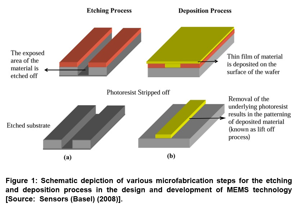
Conventional microfabrication techniques that are used in microelectronics and chip making industry can be leveraged in the field of BioMEMS for manipulating liquids and biological entities at small length scales. Figure 1 shows the microfabrication process that consists of etching and vacuum deposition of thin films on the wafer. It uses reactive ion etching along with sputter etching of the exposed area of the material for removal. Vacuum deposition process is employed to coat a thin layer of material on the surface of the wafer. Several deposition methods can be employed that include chemical vapor deposition processes process, plasma enhanced chemical vapor deposition and molecular beam epitaxial growth, which is a relatively new technique that allows deposition of films with molecular thickness [8].
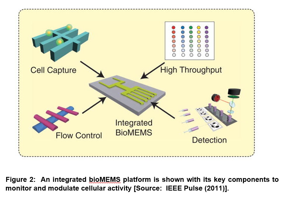
Researchers have shown that cell seeding, cell culture, modulation, monitoring, and analysis can be achieved on a single chip with high efficiency by employing miniaturization that allows the integration of various components with different functions on a bioMEMS technology platform (Figure 2). High-throughput platforms can be realized to build an array of microscale wells that are interconnected via fluidic channels. Microfabrication techniques can be leveraged to achieve such highly integrated arrays with fluidic control for individual wells [7].
A micropipette is used in the conventional cell culture approach. This is done to add the metered biochemical agents/products. This is a complex operation that produces a decreasing stimulation profile due to consumption of factors by the cell or via noncellular chemical degradation [7]. However, microfluidics technology is employed in a biochemical MEMS based cell culture and modulation of cell behavior. This approach enables the critical ability to perfuse cultured cells with a well-defined stimulus pattern. This results in facilitating stimulus–response analysis that can be used to simulate nutrient concentrations in blood following fasting and feeding [7].
Further, in the traditional method based on electrophysiology, cells are electrically induced and tracked to study how a network of neurons collectively executes high-level functions that include memory and cognition. However, individual stimulation of neuron cells is a challenge. The bioMEMS approach overcomes this problem. In bioMEMS platform, micro-patterned electrodes that are based on microelectronics fabrication process on culture platforms can be combined with microfluidics. This can allow precise temporal and spatial exposure to biochemicals that can stimulate single neurons [7]. Optical mode of bioMEMS is an advanced version of the technology that takes advantage of the photonic structures and diodes. In this design, the integrated microoptofluidic systems can be combined with the techniques of high-content imaging that enable a variety of on-chip measurements. This can also create highly portable devices that can be used in remote settings. BioMEMS mode can be employed in mechanical perturbation. This can be applied to study cell response in various ways ranging from changes in morphology to gene expression. Microfluidics technology enables such bioMEMS mechanical mode for applying quantifiable fluidic shear forces on cells [7, 8].
- BioMEMS for Early Detection and Monitoring of Cardiovascular Disease
Cardiovascular disease (CVD) affects the heart, veins, and arteries. CVD is a serious medical condition that causes heart attacks/coronary heart disease, strokes/cerebrovascular, peripheral arterial disease, rheumatic heart and congenital heart diseases as well as deep vein thrombosis and pulmonary embolisms. The fatalities that result from CVD are known to be quite devastating. Researchers have estimated that by 2030, more than 23.6 million people worldwide will die from CVD alone, which is a frightening scenario [9]. BioMEMS technology is considered very promising for early detection and effective monitoring of heart disease. It has been shown that one of the principle origins of heart failure is due to the increased pro- and anti-inflammatory cytokine levels. Researchers developed BioMEMS device based on silicon substrate for multiple cytokine detection. The fabricated BioMEMS consisted of eight gold working electrodes for the simultaneous detection of different cytokines. This was achieved by employing electrically addressable diazonium-functionalized antibodies [10].
Current research efforts are focused on developing innovative bioMEMS technologies for remote monitoring of heart failure patients. The goal of this research is to develop technologies for early detection of any medical conditions related to CVD. To this end, one novel idea is based on implementing wireless bioMEMS technology. This is for the purpose of implantation in the pulmonary artery using a minimally invasive procedure, which can subsequently measures and transmits data. It is believed that such a bioMEMS based detection platform would allow for early interventions before the situations can worsen in all CVD related medical conditions [11-14].
- BioMEMS Chip for Point-of-Care Clinical Applications
Recent studies have suggested that BioMEMS can make a significant contribution in the areas of point-of-care (POC) patient clinical care as well as the affordability of providing that care to the patients. To this end, BioMEMS platform for POC clinical applications has been considered in the case of non-communicable diseases. These include detection of protein or deoxyribonucleic acid (DNA) cancer biomarkers from serum along with the detection of micro-ribonucleic acid (micro-RNA) for cancer detection and epigenetic analysis. POC clinical applications can also be extended to the collection of exosomes and collection of circulating tumor cells. POC clinical applications of bioMEMS chip are also suggested for infectious diseases. It is crucial to obtain accurate helper T cell and viral load counts at regular intervals to monitor the health of virus-infected patient’s immune system. BioMEMS based rapid, POC detection of these infectious diseases offers promise and open up new therapeutic pathways to better manage these diseases [15].
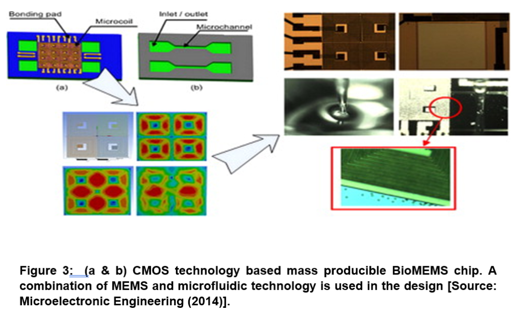
With respect to the fabrication of bioMEMS chip, researchers have leveraged complementary metal oxide semiconductor (CMOS) circuits and technology that are employed in microelectronics fabrication process. They demonstrated bioMEMS chip based on planar microcoil array as both magnetic field source and the front-end inductive sensor. This bioMEMS technology was demonstrated for highly efficient magnetic beads manipulation and a quantitative detection in point-of-care diagnostics Figure 3 [16].
- BioMEMS for Specific Drug Delivery to Enhance the Efficacy of Treatments
In recent years, extensive research and developments have taken place to develop innovative drug delivery devices that have revolutionized the course of therapeutic treatment to combat complex diseases. It has been shown that these drug delivery devices have the potential to overcome the challenges of systemic administration that limits providing site-specific high drug potency especially at the body tissues that are infected. In view of the unprecedented potentials of new drug delivery systems especially that are based on bioMEMS platform, the current research efforts in the pharmaceutical industry are directed to exploring the reliable actuating mechanisms that seek to precisely control the dispensing of drugs. The overall goal of this research is to develop a process that provides therapy and dispense drugs precisely at the infected sites. This eventually controls therapeutic effects that result in minimum toxicity. An innovative concept is based on the wireless actuation of drug delivery devices. This has been considered by the researchers lately, to adopt an intervening noninvasive approach that enables easy release of encapsulated drug compounds. Subsequently, the device is swallowed or injected and traverses through the body to reach to the desired location or specific tissue sites. A schematic illustration of the whole process is depicted in Figure 4 [17].
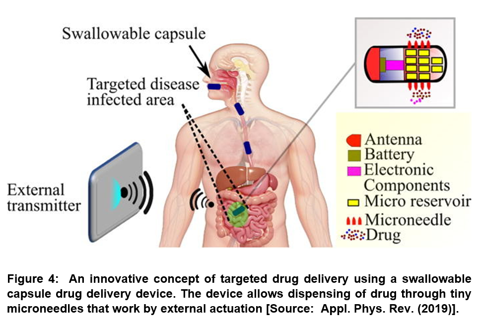
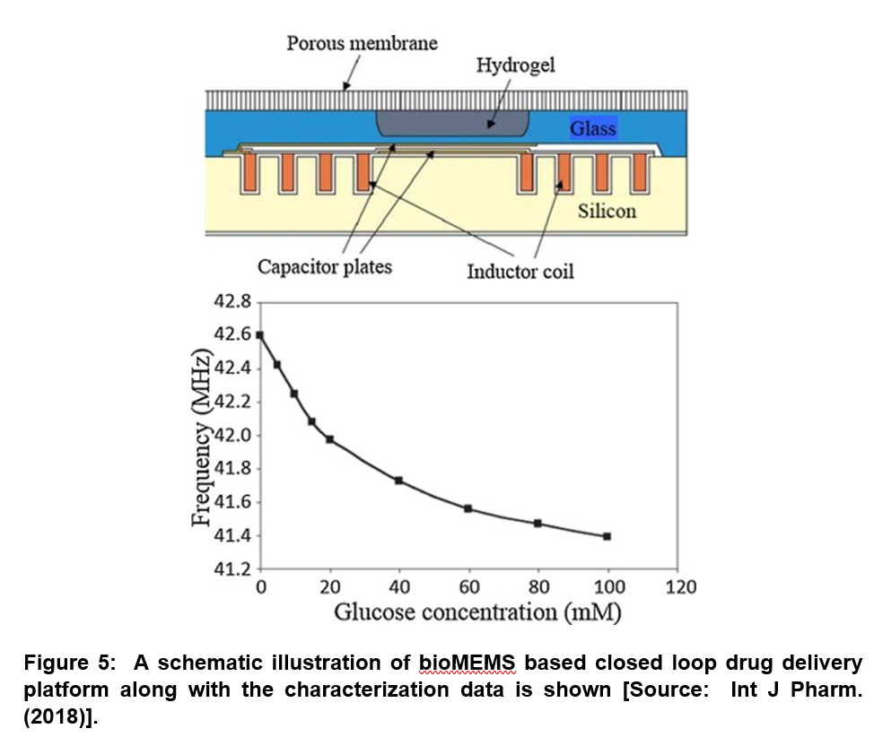
As Figure 4 shows, the efficacy of pharmaceutical treatments can be greatly enhanced by physiological feedback from the patient using the bioMEMS based biosensors platform. In related studies, researchers demonstrated closed loop drug delivery by integrating of these systems with bioMEMS based drug delivery devices (Figure 5) [18-20].
Concluding Remarks
The BioMEMS technology offers new possibilities in expanding the horizons and scope of ever growing drug delivery platforms that can impact a number of key areas in pharmaceutical and biotechnology industries. Recent studies have shown promise of bioMEMS technology platform for new innovative ways of modulating, monitoring, and accommodating biological entities. These novel techniques can be leveraged to create a more physiologically relevant environment to achieve realistic responses from cultured cells, which promotes new devices for life saving applications.
The BioMEMS field is growing rapidly, and there is a need for simplifying and standardizing BioMEMS tools. Seamless operations of BioMEMS devices are limited by two of the major obstacles that include complexity and unreliability. In future studies, we anticipate testing of new proof-of-concept bioMEMS devices for the reliability in operating these technologically important BioMEMS devices for real-world applications.
Acknowledgement
Financial assistance by Marie Skłodowska-CurieEuropean Actions Invidvidual Fellowship is thankfully acknowledged by PT.
References
[1] Bashir Rashid, BioMEMS: state-of-the-art in detection, opportunities and prospects,
Advanced Drug Delivery Reviews, 2004, 56, 1565-1586, doi: https://doi.org/10.1016/j.addr.2004.03.002.
[2] Borenstein JT, Vunjak-Novakovic G. Engineering tissue with BioMEMS. IEEE Pulse. 2011, 2, 28-34, doi: https://doi.org/10.1109/MPUL.2011.942764.
[3] Borenstein, J.T., Tupper, M.M., Mack, P.J. et al. Functional endothelialized microvascular networks with circular cross-sections in a tissue culture substrate. Biomed Microdevices, 2010, 12, 71–79, doi: https://doi.org/10.1007/s10544-009-9361-1.
[4] Menon, K., Joy, R.A., Sood, N. et al. The Applications of BioMEMS in Diagnosis, Cell Biology, and Therapy: A Review. BioNanoSci. 2013, 3, 356–366, doi: https://doi.org/10.1007/s12668-013-0112-7.
[5] Zhang, Z.; Tang, Z.; Liu, W.; Zhang, H.; Lu, Y.; Wang, Y.; Pang, W.; Zhang, H.; Duan, X. Acoustically Triggered Disassembly of Multilayered Polyelectrolyte Thin Films through Gigahertz Resonators for Controlled Drug Release Applications. Micromachines 2016, 7, 194, doi: https://doi.org/10.3390/mi7110194.
[6] Çağlayan Z, Demircan Yalçın Y, Külah H. A Prominent Cell Manipulation Technique in BioMEMS: Dielectrophoresis. Micromachines (Basel). 2020, 11(11), 990. doi: https://doi.org/10.3390/mi11110990.
[7] Erkin Seker, Jong Hwan Sung, Michael L. Shuler, and Martin L. Yarmush, Solving medical problems with BioMEMS. IEEE Pulse 2011, 2(6), 51–59, doi: https://doi.org/10.1109/MPUL.2011.942928.
[8] James T, Mannoor MS, Ivanov DV, BioMEMS – Advancing the Frontiers of Medicine, Sensors (Basel) 2008, 8, 6077-6107, doi: https://doi.org/10.3390/s8096077.
[9] Gonzalez Ivonne S and La Belle TJ; The Development of an At-Risk Biosensor for Cardiovascular Disease, Biosensors Journal 2012, 1, doi:https://doi.org/10.4303/bj/235493.
[10] Baraketa A.; Leea M.; Zinea N.; Giovanna Trivellab M.; Zabalac M.; Bausellsc J.; Sigauda M.; Jaffrezic-Renaulta N.; and Errachid A; A Fully Integrated Electrochemical BioMEMS Fabrication Process for Cytokine Detection: Application for Heart Failure. Procedia Engineering 2014, 87, 377–379, doi: https://doi.org/10.1016/j.proeng.2014.11.737.
[11] Pour-Ghaz I.; Hana D.; Raja J.; Ibebuogu UN.; Khouzam RN; CardioMEMS: where we are and where can we go?. Ann Transl Med. 2019, 7(17), 418, doi: https://doi.org/10.21037/atm.2019.07.53.
[12] Kaisti M.; Panula T.; Leppänen J; et al. Clinical assessment of a non-invasive wearable MEMS pressure sensor array for monitoring of arterial pulse waveform, heart rate and detection of atrial fibrillation. npj Digit. Med. 2019, 2, 39, doi: https://doi.org/10.1038/s41746-019-0117-x.
[13] Brugts JJ.; Radhoe SP.; Aydin D.; Theuns DA.; Veenis JF; Clinical Update of the Latest Evidence for CardioMEMS Pulmonary Artery Pressure Monitoring in Patients with Chronic Heart Failure: A Promising System for Remote Heart Failure Care. Sensors (Basel). 2021, 21(7), 2335. doi: https://doi.org/10.3390/s21072335.
[14] Abraham J.; McCann P.J.; Guglin M.E; et al. Management of the Patient with Heart Failure and an Implantable Pulmonary Artery Hemodynamic Sensor. Curr Cardiovasc Risk Rep 2020,14, 12, doi: https://doi.org/10.1007/s12170-020-00646-4.
[15] Karsten SL.; Tarhan MC.; Kudo LC.; Collard D.; Fujita H; Point-of-care (POC) devices by means of advanced MEMS. Talanta. 2015, 145, 55-59. doi: https://doi.org/10.1016/j.talanta.2015.04.032.
[16] Zheng Y.; Mannai A.; Sawan M.; A BioMEMS chip with integrated micro electromagnet array towards bio-particles manipulation. Microelectronic Engineering 2014, 128, 1, doi: https://doi.org/10.1016/j.mee.2014.06.006.
[17] Khan AN.; Ermakov A.; Sukhorukov G.; and Yang Hao; Radio frequency controlled wireless drug delivery devices, Appl. Phys. Rev. 2019, 6, 041301 doi: https://doi.org/10.1063/1.5099128.
[18] Coffel J, Nuxoll E. BioMEMS for biosensors and closed-loop drug delivery. Int J Pharm. 2018, 544(2), 335-349, doi: https://doi.org/10.1016/j.ijpharm.2018.01.030.
[19] Cobo A.; Sheybani R.; and Ellis Meng; MEMS: Enabled Drug Delivery Systems, Adv. Healthcare Mater. 2015, 4(7), 969-82, doi: https://doi.org/10.1002/adhm.201400772.
[20] Kulinsky L.; Madou M.J.; 9-BioMEMs for drug delivery applications, Editor(s): Shekhar Bhansali, Abhay Vasudev, In Woodhead Publishing Series in Biomaterials, MEMS for Biomedical Applications, Woodhead Publishing, 2012, 218-268, ISBN 9780857091291, doi: https://doi.org/10.1533/9780857096272.3.218.