The Emergence of Medical Laser based Diagnostics to Battle against Complex Diseases
Pradeep Kumar Singh1* and Shweta Kushmakar Singh2
1Department of Microbiology, S.M.M.H. Medical College, Saharanpur, UP, India
2Department of Pediatrics, All India institute of medical sciences, New Delhi, India.
Abstract
Lately, we have witnessed an explosive growth of innovative lasers and optical devices for clinical diagnostics and therapeutics including assessing human health and treating complex diseases. It is believed that light and optical techniques can have profound impacts on modern medicine. To this end, advances in medical lasers enabled biomedical optics have resulted in sophisticated technologies that integrate several other technologies including photonics with nanotechnology, biomaterials and genetic engineering, to name a few. Especially, Novel biomedical laser applications based on new laser types or novel energy delivery systems could increase the usable range of laser-tissue interactions, and thus improve target-oriented, high precision application of laser radiation in clinical practice. Here, we have presented a perspective analysis of recent demonstrations of medical lasers for complex diagnostics including clinical diagnostics applications.
Keywords: Medical laser, diagnostics, Alzheimer’s disease, skin cancer, pathogens
* Corresponding author: Dr. Pradeep Kumar Singh
E-mail address: pksingh1976a@gmail.com
DOI: https://doi.org/10.37756/bk.22.4.4.1
Article type: Perspective
Received: March 2, 2022
Revised: March 20, 2022
Accepted: March 22, 2022
Please cite this article as: Singh PK, The Emergence of Medical Laser based Diagnostics to Battle against Complex Diseases, Biotechnol. kiosk, Vol 4, Issue 4, PP: 1-8 (2022); DOI: https://doi.org/10.37756/bk.22.4.4.1.
Introduction
Medical laser applications have manifold dimensions that involve advanced technologies. These applications are aimed to meet current challenges in clinical diagnostics and therapeutics to battle against complex diseases such as blood borne pathogens, skin cancer and cardiovascular and neurodegenerative diseases, to name a few. Medical laser based diagnostic technologies are capable of addressing a wide range of health care issues that could have significant impact in the future on large sectors of the population. With respect to recent research activities and ongoing innovative developments, researchers have shown short- to mid-term improvements of both clinical diagnostics and therapeutic procedures as well as for the extension of the applicability of established techniques for new medical indications. For example, it has been shown that novel biomedical laser applications based on new laser types or innovative energy delivery systems could increase the usable range of laser-tissue interactions that could improve target-oriented, high precision application of laser radiation in clinical practice. Studies have indicated that these new energy delivery techniques together with innovative key medical techniques that are now at the translational stage into the clinics could pave the way to many future breakthroughs in medical diagnostics [1-4].
With respect to clinical applications, researchers have shown the promise of optical biopsy and non-destructive diagnosis techniques. However, these techniques still have to overcome challenges to be recognized as standard techniques for commercial applications. While histopathology of excised tissue samples is still considered the gold standard for diagnosis, latest research has focused on developing new non-invasive laser and photonics-based diagnostic techniques. A significant focus has been on oncology to develop laser based techniques that rely on the physical and biochemical changes that precede or mirror malignant changes within tissue and involve simple optical tissue interrogation techniques. Several studies have shown their accuracy in terms of sensitivity and specificity that have demonstrated the potential for a cost-effective, real-time, in-situ diagnosis for specific diseases and conditions [1, 5-13].
Here, we will take a look at some of the notable applications of laser based diagnostic tools that have been employed to combat complex medical conditions.
Detection of Pathogens in Blood Borne Infections
It is a known fact that the ability to detect and identify pathogens in blood borne infections is very important for either patient treatment/monitoring and also to make sure the safety of the donor blood supply. The existing diagnostic methodology for the detection of an infection and the identification of the responsible organism typically requires up to 72 hours depending on the pathogen. This is also dependent on the use of highly skilled personnel along with complex sample preparation. Additionally, it often requires transport of a blood sample to a microbiology laboratory for analysis. To overcome these challenges, the ability to rapidly diagnose blood borne infections on-site that does not require simple sample preparation and also highly skilled personnel is currently needed. It is believed that rapid detection would greatly enhance the ability to identify, contain, and treat blood borne infections. This can also greatly reduce the time needed to screen donated blood for infections. Moreover, the development of a blood diagnostic with these capabilities is considered a significant advancement that can enable rapid screening of patients for infections and treatments immediately. To this end, researchers have demonstrated point-of-care diagnostic instrumentation that is based on the analysis of spectra collected from a series of laser sparks formed on a blood sample. The methodology is known as Laser-Induced Breakdown Spectroscopy (LIBS), which is an established spectrochemical analysis technique. In this process, a laser pulse is used to simultaneously vaporize a small sample mass and excite the resulting atoms to emit light via formation of a hot plasma on the sample surface. Subsequently, light from the plasma is collected, spectrally resolved and finally recorded. Further, the instrument has been shown to be programmable for multiplex detection. This allows the programming to be expanded as new algorithms are developed to detect additional infections. Therefore, it is thought that a laser-based approach can achieve sensitive, multiplex detection with minimal sample preparation and provide rapid results. These important properties together with the flexibility to add new agent detection by simply adjusting the detection programming make it a promising tool for clinical diagnosis [14].
The recent studies have focused on clinical diagnostics of newly emerging agents and to screen simultaneously for multiple infectious agents. To this end, laser-based diagnostic techniques have been demonstrated to rapidly detect the presence of parasites, bacteria and viruses in human blood. For example, researchers have successfully demonstrated differentiation of blood spiked with viruses, bacteria, or protozoan parasites. This was achieved at clinically relevant levels using six blood types (O+, O-, AB+, A+, A-, B+) from different individuals with blood samples. Spiked samples were shown to be useful to detect pathogens at low concentrations in human blood using multivariate methods on carefully controlled samples. This showed the ability to detect blood samples spiked with the bacteria S. aureus and Yersinia pseudotuberculosis (Y. pseudotuberculosis), the parasite Trypanosoma cruzi (T. cruzi) and the Human Immunodeficiency Virus (HIV-1) by analysis of LIBS spectra [14].
Diagnosis of Alzheimer’s disease using Laser-Induced Breakdown Spectroscopy and Machine Learning
Alzheimer’s disease (AD) is known as a progressive incurable neurodegenerative disease and considered a major health condition in aging population worldwide. The magnitude of devastation caused by AD can be understood by an estimate that the number of AD cases in the US alone will exceed 20 million by the year 2050. It is believed that the related healthcare costs will reach $1 trillion. It is widely recognized that an early diagnosis that allows for non-invasive cost-effective tests for AD along with clear discrimination of AD from other dementias is pivotal for an efficient AD management. This is vital for the success of clinical trials of drug candidates. Recently, researchers proposed a minimally invasive approach to medical diagnosis based on LIBS liquid biopsy technique. They employed easily harvested biological fluids, such as blood, urine or saliva that were deposited and dried on solid substrates prior to laser irradiation (Figure 1) [15].
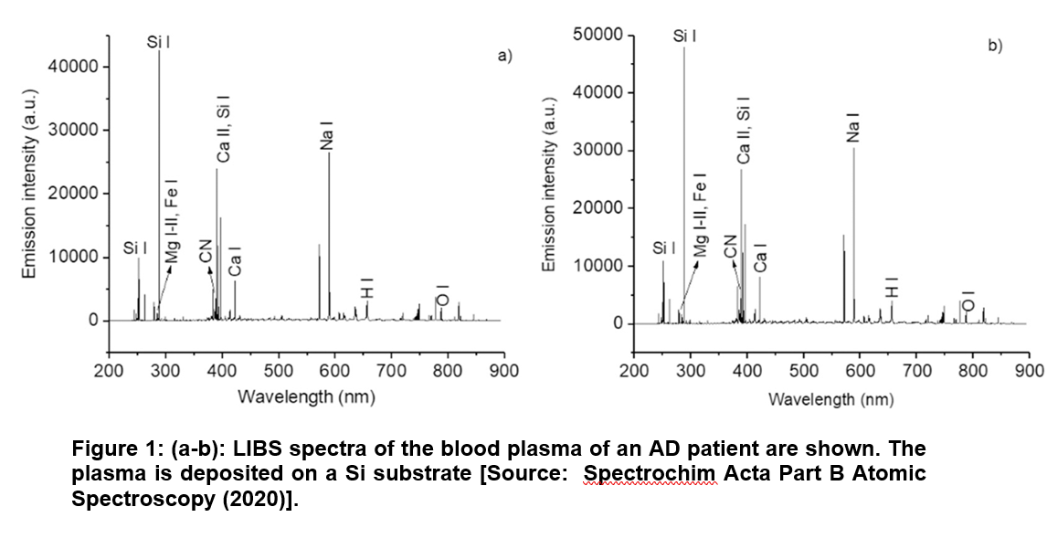
In this study, researchers proposed the combined use of LIBS and machine learning for the diagnosis of AD using biomedical fluids. They analyzed micro-drops of plasma from a cohort of AD patients and HC and focused on distinguishing the two classes with a data analysis approach based on the use of LIBS difference spectra. This study paves the way to further investigations on the potential of the combined use of LIBS and multivariate statistical approaches to analyze rapidly and relatively simply a large number of biomedical samples for the diagnosis of asymptomatic diseases [15].
Optical Coherence Tomography and Confocal Laser Scanning Microscopy: Non-Invasive Imaging for in-vivo Analysis to Combat Skin Cancer
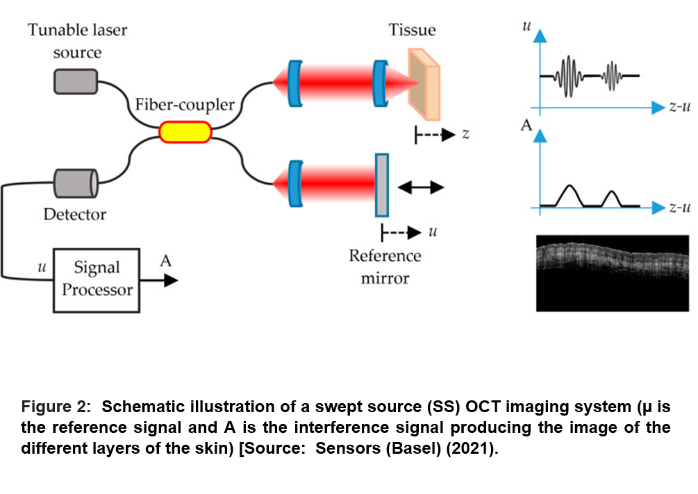
Optical Coherence Tomography (OCT) is considered a non-invasive imaging technique with an NIR laser source. OCT is employed for in-vivo skin analysis that offers real-time, cross-sectional evaluations of the skin. In the OCT process, the backscattered light from a given depth in the tissue is recombined with a static reference signal. This is followed by an interference occurs in the event of matching of both path lengths within the so-called coherence length of the light source. This enables create both two-dimensional (2D) and three-dimensional (3D) images (Figure 2) [16].
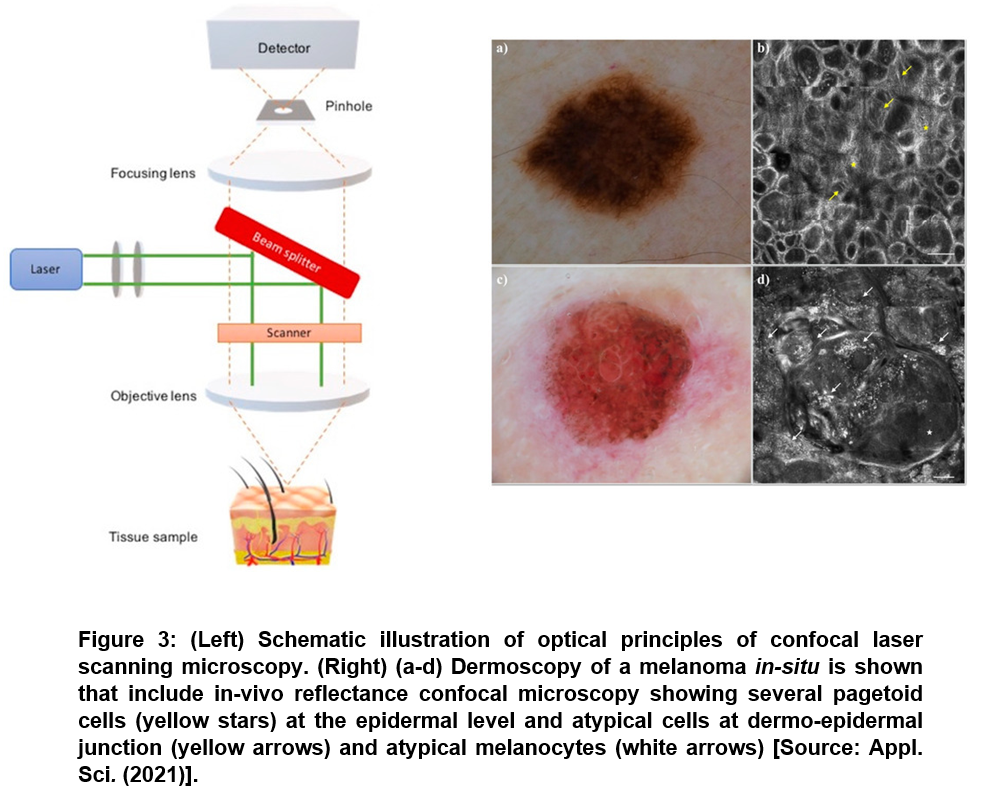
 OCT angiography (OCTA) is usually employed for the visualization of vascular patterns in the skin angiogenesis (a sign of growth and spread of cancers) that is considered helpful in improving diagnostic accuracy [17]. Another emerging technique is the confocal laser scanning microscopy (CLSM), which is a promising in-vivo and ex-vivo technique for diagnostics in clinical settings. CLSM enables a quasi-histologic view of a given tissue and that does not require a biopsy. The technique is based on refractive index of cell structures that serve as endogenous chromophores, which allows reaching a depth of exploration of 200 μm [18]. In a recent study, researchers demonstrated in-vivo CLSM in reflectance and fluorescent mode for real-time pathological examination of freshly excised specimens for diagnostic purposes. This allowed for the evaluation of margin clearance after excision in Mohs surgery (Figure 3) [18].
Conclusion
Laser based diagnostic tools have proven to be very effective in the early detection and intervention in several complex diseases. Several techniques have been shown in recent studies that offer hope to combat a number of diseases that include blood borne pathogens, neurodegenerative diseases and skin cancer, to name a few. We anticipate further development of laser diagnosis in the near future for non-invasive and early detection of diseases for timely intervention and cure.
References
[1] Ronald S and Lothar L, Laser-advanced new methods for diagnostics and therapeutics. Photonics & Lasers in Medicine 2016, 5 (1):1-4, DOI: https://doi.org/10.1515/plm-2015-0046.
[2] Geoghegan S, Lasers in medical diagnosis and therapy by Stephan Wieneke and Christoph Gerhard. Australas Phys Eng Sci Med 2019, 42:899–900, DOI: https://doi.org/10.1007/s13246-019-00777-y.
[3] Coda S, Siersema PD, Stamp GW, Thillainayagam AV, Biophotonic endoscopy: a review of clinical research techniques for optical imaging and sensing of early gastrointestinal cancer. Endosc Int Open 2015, 3(5):E380–92, DOI: https:doi.org/10.1055/s-0034-1392513.
[4] Sroka R, Stepp H, Hennig G, Brittenham GM, Rühm A, Lilge L, Medical laser application: translation into the clinics. J Biomed Opt 2015, 20(6):061110, DOI: https://doi.org/10.1117/1.JBO.20.6.061110.
[5] Yun SH, Kwok SJJ, Light in diagnosis, therapy and surgery. Nat Biomed Eng 2017, 1:0008, DOI: https://doi.org/10.1038/s41551-016-0008.
[6] Srinivasan R, Ablation of polymers and biological tissue by ultraviolet lasers. Science 1986, 234:559–565, DOI: https://doi.org/10.1126/science.3764428.
[7] Jacques SL, Optical properties of biological tissues: a review, Phys Med Biol 2013, 58:R37–61, DOI: https://doi.org/10.1088/0031-9155/58/11/R37.
[8] Humar M, Yun SH, Intracellular microlasers, Nat Photonics 2015, 9:572–576, DOI: https://doi.org/10.1038/nphoton.2015.129.
[9] Kut C., et al., Detection of human brain cancer infiltration ex vivo and in vivo using quantitative optical coherence tomography, Sci Transl Med 2015, 7:292ra100, DOI: https://doi.org/10.1126/scitranslmed.3010611.
[10] Friedman Aa, Letai A., Fisher DE., Flaherty KT., Precision medicine for cancer with next-generation functional diagnostics, 2015, Nat Rev Cancer, 15:747–756, DOI: https://doi.org/10.1038/nrc4015.
[11] Ming K, Kim J, Biondi MJ, Syed A, Chen K, Lam A, Ostrowski M., Rebbapragada A, Feld JJ, Chan WC, Integrated quantum dot barcode smartphone optical device for wireless multiplexed diagnosis of infected patients. ACS Nano 2015, 9(3):3060-74, DOI: https://doi.org/10.1021/nn5072792.
[12] Pritzker KPH, & Nieminen HJ, Needle Biopsy Adequacy in the Era of Precision Medicine and Value-Based Health Care. Arch Pathol Lab Med 2019, 143:1399–1415, DOI: https://doi.org/10.5858/arpa.2018-0463-RA.
[13] Hollon TC, Pandian B, Adapa AR, Urias E, Save AV, Khalsa SSS, Eichberg DG, Amico RSD, Farooq ZU, Lewis S, Petridis PD, Marie T, Shah AH, Garton HJL, Maher CO, Heth JA, Mckean EL, Sullivan SE, Hervey-jumper SL, … Orringer DA, Near real-time intraoperative brain tumor diagnosis using stimulated Raman histology and deep neural networks. Nat Med 2020, 26:52–58, DOI: https://doi.org/10.1038/s41591-019-0715-9.
[14] Multari RA, Cremers DA, Nelson A, Karimi Z, Young S, Fisher C, Duncan R, The use of laser-based diagnostics for the rapid identification of infectious agents in human blood, J Appl Microbiol 2019, 126(5):1606-1617, DOI: https://doi.org/10.1111/jam.14222.
[15] Gaudiuso R, Ewusi-Annan E, Xia W, Melikechi N, Diagnosis of Alzheimer’s disease using laser-induced breakdown spectroscopy and machine learning. Spectrochim Acta Part B Atomic Spectroscopy. 2020, 171:105931, DOI: https://doi.org/10.1016/j.sab.2020.105931.
[16] Rey-Barroso L, Peña-Gutiérrez S, Yáñez C, Burgos-Fernández FJ, Vilaseca M, Royo S, Optical Technologies for the Improvement of Skin Cancer Diagnosis: A Review. Sensors (Basel) 2021, 2;21(1):252, DOI: https://doi.org/10.3390/s21010252.
[17] Deegan AJ, Talebi-Liasi F, Song S, Li Y, Xu J, Men S, Shinohara MM, Flowers ME, Lee SJ, Wang RK, Optical coherence tomography angiography of normal skin and inflammatory dermatologic conditions. Lasers Surg. Med 2018, 50:183–193, DOI: https://doi.org/10.1002/lsm.22788.
[18] Guida S, Arginelli F, Farnetani F, Ciardo S, Bertoni L, Manfredini M, Zerbinati N, Longo C, Pellacani G, Clinical Applications of In Vivo and Ex Vivo Confocal Microscopy. Appl. Sci 2021, 11:1979, DOI: https://doi.org/10.3390/app11051979.
Rights and Permissions
Open Access This article is licensed under a Creative Commons Attribution 4.0 International License, which permits use, sharing, adaptation, distribution and reproduction in any medium or format, as long as you give appropriate credit to the original author(s) and the source, provide a link to the Creative Commons license, and indicate if changes were made. The images or other third credit line to the permitted by statutory regulation or exceeds the permitted use, you will need to obtain permission directly from the copyright holder. To view a copy of this license, visit http://creativecommons.org/licenses/by/4.0/
Creative Commons This is an open access article distributed under the terms of the Creative Commons CC BY license, which permits unrestricted use, distribution, and reproduction in any medium, provided the original work is properly cited. You are not required to obtain permission to reuse this article. To request permission for a type of use not listed, please contact Biotechnology Kiosk.